Liver - sinusoids (100x objective lens)


This image shows hepatocytes (He) and sinusoids (Si) at high magnification. The hepatocytes are polygonal in shape with a circular nucleus (N) that is rimmed by heterochromatin (Hc) and often contains a prominent nucleolus (Nu). Hepatocytes are attached to several adjacent hepatocytes to for a plates or trabecula that is two cells thick and the hepatocytes are so arranged that every cell is exposed on several sides to the sinusoids. The dark lines that can be seen between some cells delineate the cell boundary (CB) where two cells are attached. The sinusoidal lining cells (SLC) that line the irregularly shaped sinusoids have flattened, darkly stained nuclei that may bulge into the lumen of the sinusoids. There are two types of sinusoidal lining cells cells: endothelial cells and Kupffer cells. The endothelial cells form a discontinuous simple squamous epithelium that allows the fluid components of the blood to enter the space of Disse (SD) located between the surface lining (SL), formed by the thin cell bodies of the endothelial cells, and the apical surfaces of the hepatocytes. Kupffer cells are macrophages that become part of the sinusoidal lining and phagocytose particulate matter from the blood as it flows through the sinusoids. These two cells types can not be easily distinguished on the basis of morphology alone. Only when the Kupffer cells are filled with phagocytosed particles, either natural or artificial, can they be differentiated from endothelial cells. Both red blood cells (RBC) and white blood (WBC) cells can be seen in the sinusoids.

Stain = H&E


Go to lower magnification Go 
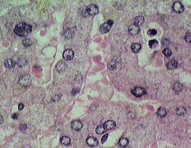
to another high magnification image of sinusoids Return to Digestive System. Return to the Table of Contents.

Show Labels


Copyright by: Paul B. Bell, Jr. & Barbara Safiejko-Mroczka

The University of Oklahoma

Version: 001206