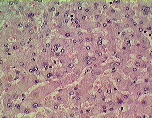
This image shows the
anastomosing plates of hepatocytes (He) that
make up the bulk of the liver. Hepatocytes are polygonal in shape with
a circular nucleus
(N). The nucleus is usually outlined by a dark
circle of heterochromatin (Hc),located
just beneath the nuclear envelope, and often contains a prominent nucleolus
(Nu).
Hepatocytes are arranged in plates or trabeculae that are two cells thick
and the hepatocytes are so arranged that every cell is both attached to several
of its neighbors and also exposed on several sides to blood filled sinusoids
(Si). The irregularly shaped sinusoids, containing red
blood cells (RBC), are lined by one of two types of cells: endothelial
cells and Kupffer cells. Both types of cell shave flattened nuclei and they
are identified here simply as sinusoidal
lining cells (SLC) because they
are difficult to tell apart. There
is narrow space between the lining cells and the surfaces of the hepatocytes,
called the Space of Disse (SD).