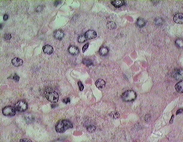
Liver - sinusoids (100x objective lens)


This image shows several "cords" (as they are sometimes incorrectly described) of hepatocytes (He) separated by sinusoids (Si). The hepatocytes can be identified on the basis of their polygonal in shape and circular nucleus (N) that is rimmed by heterochromatin and often contains a prominent nucleolus (Nu). The sinusoidal lining cells (SLC), in contrast, have flattened, darkly stained nuclei that may bulge into the lumen of the sinusoids and, at most, a thin flange of cytoplasm that forms the lining of the sinusoids. Red blood cells are often present the sinusoids but they do not enter the space of Disse (SD) located between the surface lining and the apical surfaces of the hepatocytes.

Stain = H&E


Return to the first high magnification image of sinusoids Go to a third high magnification image of sinusoids Return to Digestive System. Return to the Table of Contents.

Show Labels


Copyright by: Paul B. Bell, Jr. & Barbara Safiejko-Mroczka

The University of Oklahoma

Version: 001206