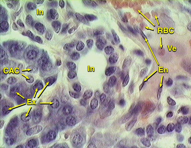
This image shows two
intercalated ducts (In), located
between acini of basophilic exocrine secretory cells
(Ex) and a vein (Ve). The wall of the
ducts is a simple epithelium of cuboidal to low cuboidal cells, surrounded
by a thin layer of connective tissue. Centroacinar cells
(CAC) can be distinguished from the surrounding basophilic cells by
their unstained cytoplasm. The vein is lined by endothelial
cells (En) and contains red blood cells RBC).