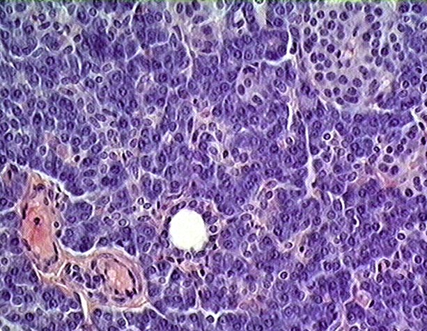
Pancreas - intercalated duct (40x objective lens)


This image of the pancreas shows a cross sectioned intercalated duct (In), composed of a simple epithelium of cuboidal to low columnar cells. Above and to the left is a smaller intercalated duct (In) cut tangentially such that the nuclei of the epithelial cells can be seen but not the lumen. Surrounding the duct are numerous acini of basophilic exocrine secretory cells and several islands of light staining endocrine cells, the islets of Langerhans (IL). An arteriole (Ar) and a vein (V) are located in the interlobular connective tissue (CT), which forms as an expansion of the septa (Se).
Stain = H&E

Go to lower magnification Go to higher magnification of exocrine acini Go to higher magnification of another intercalated duct Return to Digestive System. Return to the Table of Contents.

Show Labels


Copyright by: Paul B. Bell, Jr. & Barbara Safiejko-Mroczka

The University of Oklahoma

Version: 010815