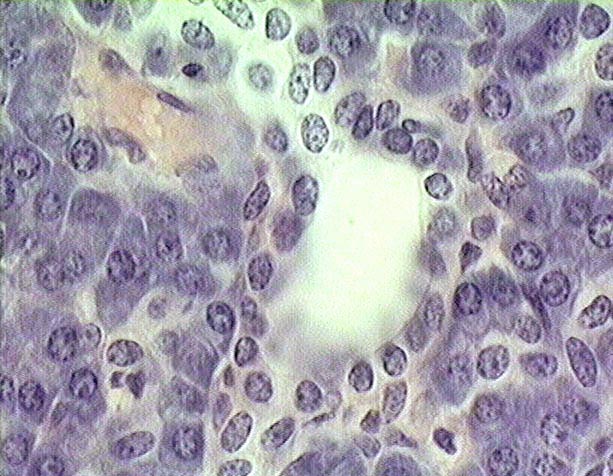
This image shows a tangential
section through an intercalated ducts (In),
located among acini of basophilic exocrine secretory cells.
The wall of the duct is a simple epithelium of cuboidal to low cuboidal cells,
surrounded by a thin layer of connective tissue. A blood
vessel (BV), probably a smal vein, lined by endothelial cells is seen in
the upper left quadrant of the image.