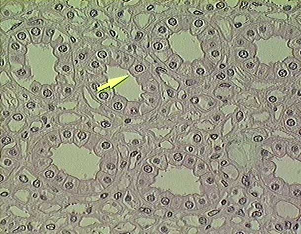
(2) thin segments of the loops of Henle. The walls are composed of a simple squamous epithelium with relatively thick cells with oval nuclei. The nuclei often bulge into the lumen of the tubule.
(3) straight capillaries (vasa recta) of the medullary plexus of capillaries. The walls are composed of a simple squamous epithelium with relatively thin cells with flattened nuclei. The nuclei often bulge into the lumen of the capillary.
| Go to the lower magnification image | Return to Urinary System. | Return to the Table of Contents. |
Copyright Paul B. Bell, Jr. & Barbara Safiejko-Mroczka
The University of Oklahoma
Version 010603