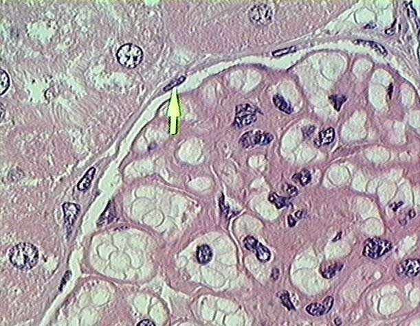
Kidney
- renal cortex - plastic section (100x objective lens)
-
This very high magnification
image shows:
-
- (1) A portion of
the renal corpuscle. The corpuscle is surrounded
by an outer layer
of simple squamous epithelium (arrow), which forms the parietal
layer of Bowman's capsule. The bulk of the corpuscle
is the glomerulus, consisting of a capillary
tuft covered by a single layer of podocytes,
which form the inner or visceral layer of Bowman's capsule.
The two layer of Bowman's capsule are separated by the urinary
space (sinus).
-
- (2) Proximal
convoluted tubules.
Note the brush border on the apical (luminal)
surfaces of the cuboidal cells the form the walls of the tubules.
- Stain =H&E.
-
Copyright
Paul B. Bell, Jr. & Barbara Safiejko-Mroczka
The
University of Oklahoma
Version 010603