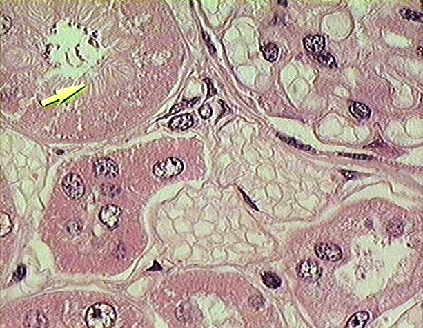
Kidney
- renal cortex -
plastic section (100x objective lens)
-
This very high magnification
view of a plastic section from the renal cortex shows:
-
- (1) a portion of
the renal corpuscle (in the upper right corner).
Note the squamous epithelial cells with flattened
nuclei that form the parietal (outer) layer of Bowman's capsule, the capillaries
of the glomerular capillary tuft, and the rounded nuclei of podocytes
that cover the capillaries to form the visceral layer of Bowman's capsule.
-
- (2) a proximal
convoluted tubule (upper left), characterized by an apical brush
border of microvilli (arrow), which reduces the internal diameter of
the tubule.
-
- (3) profiles of two
distal convoluted tubule (lower right and lower
center of the image), which lack a brush border and have a larger diameter
lumen.
(4) capillaries and veins.
Both have walls of endothelial cells with flattened nuclei. The veins have a
larger diameter and are filled with irregularly shaped erythrocytes.
- Stain = H&E.
-
Copyright
Paul B. Bell, Jr. & Barbara Safiejko-Mroczka
The
University of Oklahoma
Version 010603