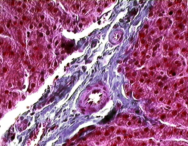
This image from the
liver of a pig showing a septum (Se) containing
two branches of the hepatic artery (HA). The
contracted appearance of these blood vessels, in which the lumen is partially
or totally occluded by the endothelial cells, is a typical postmortem artifact
of liver tissue. The large irregular spaces around and within the septum are
shrinkage artifacts (*). However,
other spaces, lined by flattened endothelial cells, are lymphatic vessels,
which drain excess fluid from the liver.