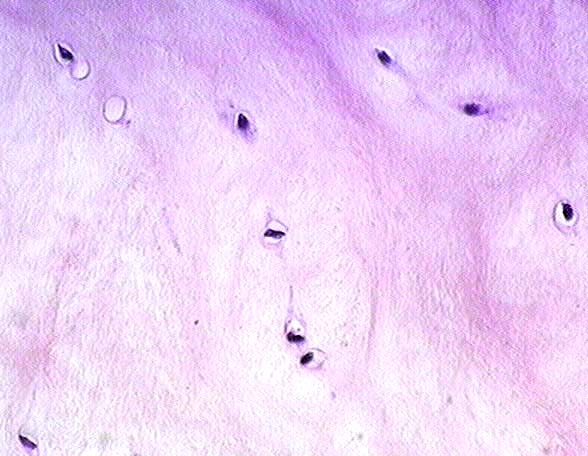
Fibrocartilage (40x objective lens)
At this magnification the eosinophilic staining of the type I collagen fiber bundles can be clearly seen.
The presence of lacunae,
occupied by
chondrocytes
, and the presence of
collagen fibers
mark this as fibrocartilage.
Stain = hematoxylin and eosin
Go to lower magnification
Go to high magnification view of hyaline cartilage
Return to Cartilage
Return to the Table of Contents
|
Copyright by:
Paul B. Bell, Jr.
&
Barbara Safiejko-Mroczka
|
The University of Oklahoma
Version: 001206