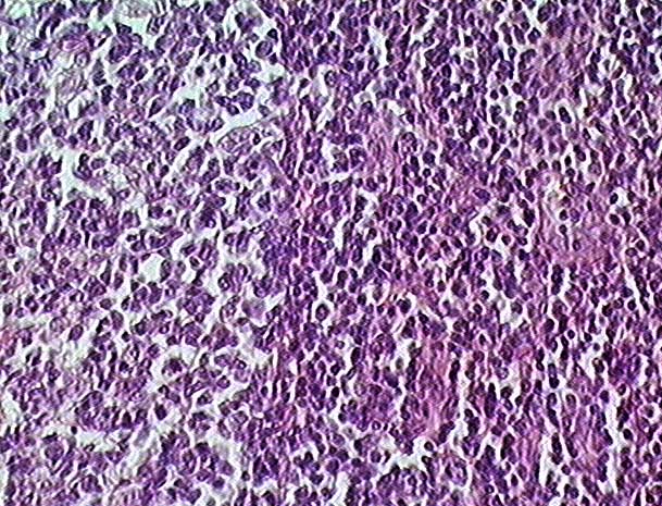
Palatine tonsil (40x objective lens)
This image shows the boundary between a germinal center, to the left, and the more densely packed cells of a lymphatic nodule, to the right. Note the higher cell density and the presence of pink-staining collagenous connective tissue in the nodule.
Stain = H&E
Go to lower magnification
Go to higher magnification
Return to Lymphatic
Return to the Table of Contents
|
Copyright by:
Paul B. Bell, Jr.
&
Barbara Safiejko-Mroczka
|
The University of Oklahoma
Version: 001206